Classification
To ensure holistic assessment and treatment of DFUs, the wound should be classified according to a validated clinical tool (Frykberg and Banks, 2015).
Classification systems have been developed to grade the severity of diabetic foot ulcers, provide prognosis on healing and aid in the formulation of treatment plans.
There are a variety of different classification systems that are used, each with their own merit.
The SINBAD (site, ischaemia, neuropathy, bacterial infection, area and depth) framework uses a scoring system that helps predict outcomes and is a simplified version of a previous classification system, (WUWHS, 2016). It is useful across multi-disciplinary teams.
SINBAD system for classifying and scoring foot ulcers
| Category | Definition WUWHS | SINBAD Score |
|---|---|---|
| Site |
|
|
| Ischaemia |
|
|
| Neuropathy |
|
|
| Bacterial infection |
|
|
| Area |
|
|
| Depth |
|
|
| Total possible score | 6 |
Adapted from North West Podiatry Services, 2014
The University of Texas Diabetic Foot Ulcer Classification System
The University of Texas system grades diabetic foot ulcers by depth and then stages them by the presence or absence of infection and ischaemia:
Grade 0 – pre-or post-ulcerative site that has healed
Grade 1 – superficial wound not involving tendon, capsule, or bone
Grade 2 – wound penetrating to tendon or capsule
Grade 3 – wound penetrating bone or joint
Within each wound grade there are four stages:
Stage A – clean wounds
Stage B – non-ischaemic infected wounds
Stage C – ischaemic non infected wounds
Stage D – ischaemic infected wounds
(Wound Educators, 2016)
The University of Texas scale is one of the most well-established systems. It represents all aspects of assessment and cross-references them alongside each other, showing a two-part score that includes grade and stage. It gives the clinician a complete picture of the individual wound (WUWHS, 2016).
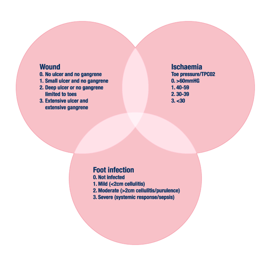
More recently, a new classification system of ‘the threatened lower limb’ WIFI (Wound, Ischaemia, Foot, Infection) has been developed for use in diabetic and non-diabetic patients. This system has been adopted by the Society for Vascular Surgery (SVC, USA; WUWHS, 2016).
Structure of the Wound/Ischaemia/Foot Infection (WIFI) System

The Wagner Scale, which assesses ulcer depth together with presence of gangrene and loss of perfusion over six grades (0-5).
NB - It does not fully take into account infection and ischaemia, (WUWHS, 2016).
Classification systems should be used consistently across the healthcare team and be recorded appropriately in the patient’s records. However, it is the assessment of the wound that informs management (Wounds International, 2013).
NICE (2015, 2019), in its Diabetic Foot Problems: prevention and management guidance, recommends the use of the SINBAD classification, or the University of Texas classification system.
Please refer to your local policy and guidelines for Managing the Diabetic Foot, for the relevant classification system.
SKIN TONES
The diabetic foot and the symptoms of ulceration can be harder to see in people who have dark skin tones. Any changes in colour need to be assessed and monitored based on that person’s baseline skin tone. The affected foot, where possible, should be compared to the other foot for comparative skin tones.
Infection diagnosis can also be challenging for those with dark skin tones, as severe diabetic foot ulcers can often present as black and or brown eschar. It is vital that a thorough skin assessment is carried out to ensure that signs and symptoms of infection, eschar etc., are not overlooked. (Wounds UK, 2021, Nair et al., 2022).
In 2014, Edmonds and Foster also described six stages of the diabetic foot to provide a framework for managing diabetic foot wounds:
| Stage | Clinical Condition |
|---|---|
| 1 | Normal |
| 2 | High risk |
| 3 | Ulcerated |
| 4 | Infected |
| 5 | Necrotic |
| 6 | The unsalvageable foot |
Table showing the six stages of the diabetic foot (Edmonds & Foster, 2014).
This system covers the whole spectrum of diabetic foot disease in six stages. It describes the natural history of each of the six clinical scenarios and emphasises the relentless progression to the end stage of necrosis.