Topic Five: Stage 5 - The Necrotic Foot
This is the end stage of the natural history of the diabetic foot. It is characterised by the presence of necrosis (gangrene) threatening the loss of the limb. Early diagnosis and intervention can save the limb. Necrosis can involve skin, subcutaneous tissue and fascial layers.
Infection is the precursor to wet necrosis, and ischaemia is the precursor to dry necrosis; both can be rapid and devastating and are termed “diabetic foot attacks”.
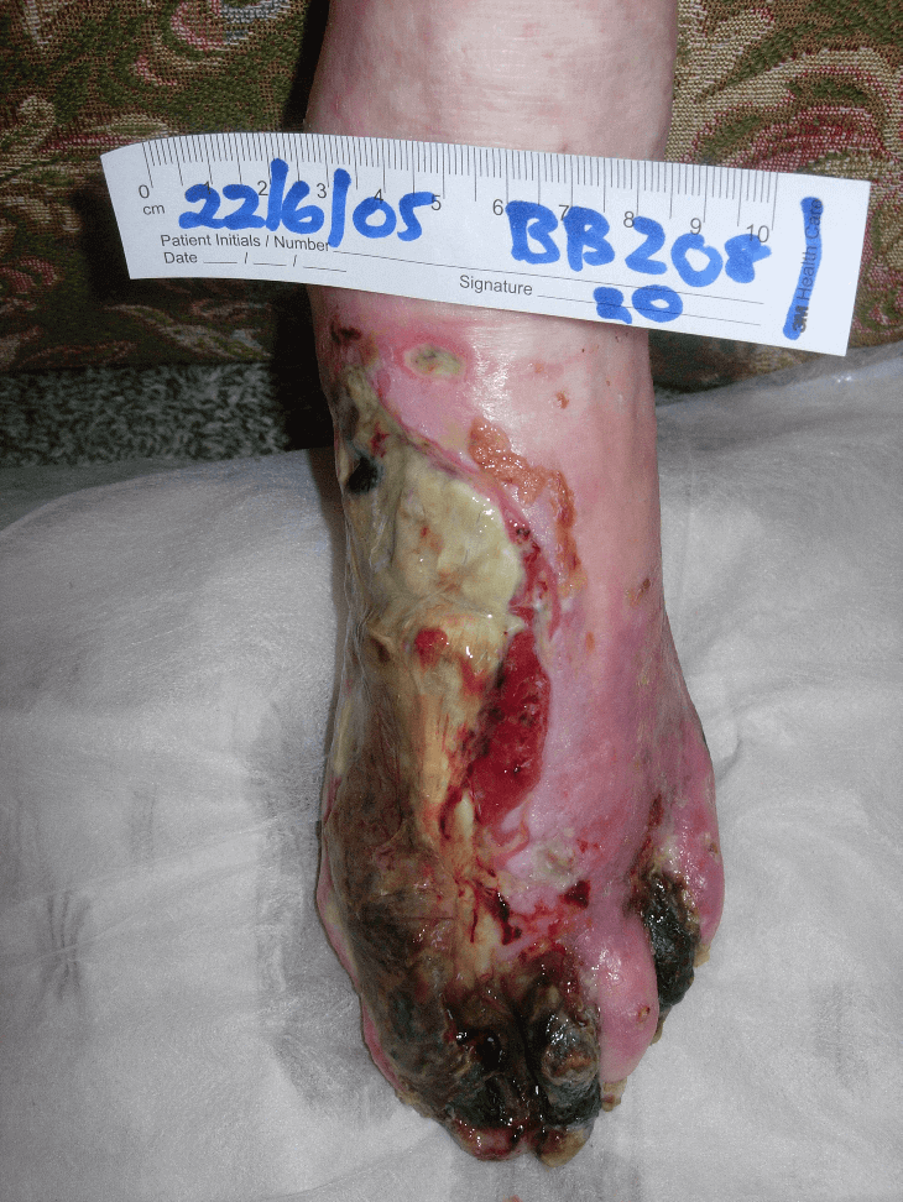
The wet necrotic foot
In wet necrosis, the tissues are grey, black, brown, white or greenish, moist and often malodorous. Adjoining tissues are infected, and pus may discharge from the ulcerated demarcation line between necrotic and viable tissue (Edmonds & Foster, 2014). Wet necrosis is secondary to septic vasculitis associated with severe soft tissue infection and ulceration and is the most common cause of necrosis in the diabetic foot.
Wet necrosis is prominent in both neuropathic and neuroischaemic foot ulcers. However, on the neuroischaemic foot reduced arterial perfusion to the foot resulting from atherosclerotic disease of the leg arteries is also a predisposing factor.
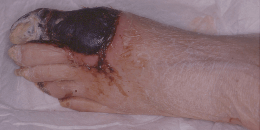
The dry necrosis
Dry necrosis is hard blackened mummified tissue and there is usually a clean demarcation line between necrosis and viable tissue.
Dry necrosis can be seen in three situations in the ischaemic foot:
- Critical ischaemia
- Acute ischaemia
- Renal ischaemia
Dry necrosis takes place gradually, progresses slowly, and affects the lower extremities of the body (toes and feet) due to insufficient blood supply to the tissues (ischaemia) and the absence of infection. It is particularly common in those suffering from arteriosclerosis, high cholesterol, smoking and diabetes.
Critical ischaemia
PVD usually progresses steadily in the diabetic patient, but a severe reduction in the arterial blood supply results in reduced perfusion to the feet, leading to a state of critical ischaemia. The final reduction in blood supply is caused by a thrombotic event. Toes may become cyanosed and progress to necrosis. Early revascularisation is crucial.
Acute ischaemia
Is caused by a thrombosis complicating an atherosclerotic stenosis in the superficial femoral or popliteal artery or by an embolus from proximal atherosclerotic plaques in the aorta, iliac femoral or popliteal arteries (Edmonds & Foster, 2014). Emboli may occur due to atrial fibrillation or myocardial infarction. Patients may present with sudden onset of pain with pallor of the foot followed by mottling and a grey discolouration.
Renal ischaemia
Both diabetic foot disease and chronic kidney disease are strong predictors for the development of diabetic foot ulceration (McIntosh & MacGilchrist, 2018). Renal diseases increase the incidence of peripheral arterial disease (PAD) and as the disease progresses so does the severity of PAD (Behan & Reyzelman, 2018).
Poor perfusion to the feet during dialysis and anaemia are some of the mechanisms by which the risk factors are increased in people with renal failure (Foster, 2017). Those with advanced renal dysfunction are at an increased risk, and in patients with both renal failure and foot complications, this is much greater (McIntosh & MacGilchrist, 2018).
MANAGEMENT
Patients should be admitted immediately for urgent investigations and multidisciplinary management. (Edmonds & Foster, 2014).
Wound control
In the neuropathic foot, operative debridement is always indicated for wet necrosis. Amputation may be necessary, this includes:
- Partial or total amputation of necrotic toes
- Ray amputation
- Transmetatarsal amputation
- Chopart or Linsfranc partial foot amputations
- Symes amputations
Post-operative wounds are treated with NPWT. In the neuropathic foot, healing should be achieved if the bioburden remains controlled.
The neuroischaemic foot is often complicated by wet necrosis and this should be removed urgently when it is associated with severe spreading sepsis.
Dry necrosis in the neuroischaemic foot, which is limited to one or two toes can sometimes be managed conservatively and auto-amputation may occur.
Vascular control
All ischaemic feet that present with necrosis must have Doppler studies followed by Duplex angiography to show stenosis or occlusions of the arteries of the leg (Edmonds & Foster, 2014). In dry necrosis, revascularisation is necessary to maintain the viability of the limb. In wet necrosis, it is necessary to heal the tissue deficit after operative debridement.
Angioplasty may be effective at restoring perfusion and achieving healing in smaller lesions. Where there is full-thickness necrosis, an arterial bypass may be necessary to optimise perfusion and the chance of saving tissue. It may take several weeks to see significant clinical improvement of the wound(s). Also, unilateral oedema may develop post-surgery, which can impede healing (Vuolo, 2009).
Microbiological control
The microbial principles of managing wet necrosis are similar to those of the management of infection in Stage 4. Patients with wet necrosis due to severe infection need immediate treatment with broad-spectrum intravenous antibiotics, even if they are apyrexial. Deep swabs and tissue specimens should be sent for microbiology, and when the results are obtained, the antibiotic regimen can be tailored to the sensitivities.
Mechanical control
All patients should remain on bed rest with elevation to relieve swelling. Heel protection and pressure area care should be maintained. If surgery is required, bed rest should continue after surgery until the ulcer is healed.
Patients’ skin should be assessed regularly, as they will be at risk of developing pressure ulcers, so should be nursed appropriately with a pressure-relieving mattress and regular repositioning.
Metabolic control
Good management of diabetes will ensure that the patient has the best chance of healing. Sliding scale insulin may be necessary, and patients may also have cardiac and renal impairment, which will require management (Vuolo, 2009).
Educational control
Patients and/or carers must be informed as investigations are carried out and management plans are made so they can fully understand the consequences.
Patients will need to understand the importance of bed rest, heel protection and pressure area care (McIntosh, 2007; Wounds UK, 2021).