Topic Four: Stage 4 - The Infected Foot

The Infected Foot
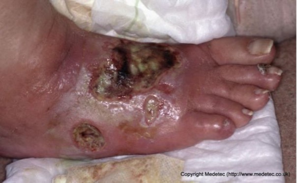
The patient now enters Stage 4 because the foot is now infected and microbial control has been lost. Infection is the main pathology that destroys most diabetic feet; therefore, infection is a medical emergency (Edmonds & Foster, 2014). Classic signs of infection may be absent or reduced due to neuropathy and/or ischaemia. Infection can spread quickly to the leg; therefore, rapid treatment is essential.
www.medetec.co.ukThe end stage of tissue death is reached quickly, and there is a limited window of opportunity for intervention. There is reduced host response to infection, particularly in diabetic patients with impaired renal and liver function.
Infection in the diabetic foot is often worse than it appears, due to the absence of the normal signs of infection. White blood cell count and body temperature may be normal even in severe infections. 50% of episodes of severe infections will provoke a fever or leucocytosis (Edmonds & Foster, 2014). However, serum CRP is a good indicator of the severity of the infection.
These infections usually take one of the following forms:
- Cellulitis – defined as an infection of skin and subcutaneous tissue and presenting as redness or an erythema
- Deep-skin and soft-tissue infections
- Acute osteomyelitis
- Chronic osteomyelitis
The following stages of diabetic foot infection may be recognised:
- Mild infection – Infection of the ulcer itself, namely local infection with minimum cellulitis >0.5 to ≤ 2 cm from the ulcer
- Moderate infection
- Severe deep infection – the most common manifestation is cellulitis
(Edmonds & Foster, 2014)
The classifications can be seen in more depth in the following table.
Infection can be classified into the presence and severity of an infection of the foot in a person with diabetes.
| Clinical classification of infection, with definitions | Classification |
|---|---|
Uninfected
|
1 - Uninfected |
Infection |
|
At least two of these items are present:
And no other cause(s) of an inflammatory response of the skin (e.g. trauma, gout, acute Charcot neuro-osteoarthropathy, fracture, thrombosis or venous stasis) Infection with no systemic manifestations (see below) involving:
|
2 - Mild infection |
Infection with no systemic manifestations, and involving:
|
3 - Moderate infection |
Any foot infection with associated systemic manifestations (of the systemic inflammatory response syndrome [SIRS]), as manifested by ≥2 of the following:
|
4 - Severe infection |
Note: Infection refers to any part of the foot, not just of a wound or an ulcer. Erythema in any direction, from the rim of the wound. The presence of clinically significant foot ischaemia makes both diagnosis and treatment of infection considerably more difficult.
Adapted from Lipsky et al., 2012; IWDF, 2019
Osteomyelitis can be a common complication of a diabetic foot infection. It is often associated with a high encumbrance of mortality. Osteomyelitis is the inflammation of bone secondary to infection, and in the diabetic foot, is mostly the consequence of a soft tissue infection. It can affect any bone, but largely the forefoot.
Osteomyelitis may be present underlying any DFU, especially those that have been present for many weeks or that are wide, deep, located over a bony prominence, showing visible bone or accompanied by an erythematous, swollen (“sausage”) toe. If underlying osteomyelitis is not determined and treated properly, the wound is unlikely to heal. Osteomyelitis can be difficult to diagnose in the early stages (Giurato et al., 2017; IWGDF, 2019)
MANAGEMENT
There are 3 crucial decisions when managing an infected foot ulcer
- Confirm infection to commence antibiotic therapy
- Select appropriate antibiotic therapy
- Consider method of debridement to remove non-viable tissue
- Sharp
- Surgical
- Biosurgical
Microbiological control
Patients with a DFU should have a rapid assessment at the earliest opportunity, preferably within one working day of presentation – or sooner in the presence of severe infection (TRIEPodD-UK, 2012; SIGN, 2010; Wounds UK, 2021).
When managing these very difficult and unstable feet, decisions made should be guided by the signs and symptoms of infection, results of wound swabs and tissue cultures and the past and present knowledge of the individual patient.
Microbiology of diabetic feet can be caused by:
- Gram-positive bacteria
- Gram-negative bacteria
- Anaerobic bacteria
- Singularly or in a combination
(Edmonds & Foster, 2014)
The antibiotic regimen will depend upon the type of ulcer (neuropathic/neuroischaemic), the degree of cellulitis and the presence, degree of/ or absence of osteomyelitis (Vuolo, 2009, IWGDF 2019).
Wound control
Debridement should be carried out to reveal the true extent of the wound in both neuropathic and neuroischaemic ulcers by autolytic, sharp or surgical debridement (Vuolo, 2009; WUWHS, 2016).
Severe infections in both neuropathic and neuro-ischaemic foot may require surgical debridement.
Vascular control
Intervention to re-perfuse the neuroischaemic foot may be required after surgical debridement, to control infection and promote wound healing. This may be achieved via angioplasty or bypass surgery.
Mechanical control
All patients with infected feet should not walk, should be on bed rest and use crutches or a wheelchair for trips to the bathroom. Heels should be protected with special heel protectors and regular, frequent turning in bed. Post-debridement off-loading may be achieved with casting techniques.
Educational control
Patients need to learn the signs and symptoms of infection and what to do once they have developed an infection.