Topic Three: Stage 3 - The Ulcerated Foot
The Ulcerated Foot
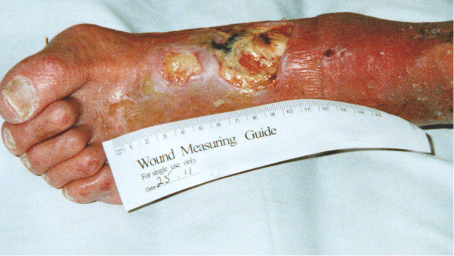
A foot reaches stage 3 when the skin has broken down and ulceration is present. Any break in the skin is a route that bacteria can enter. A foot ulcer is an important sign of systemic disease, which leads to a 50 % mortality of diabetic foot patients after five years (Edmonds & Foster, 2014).
Diabetic ulcers can rapidly deteriorate. Each ulcer should be assessed and managed individually, but it is important to differentiate between ulcers in a neuropathic foot compared with those in the neuroischaemic foot.
Neuropathic ulcer
Elevated plantar pressures are known to contribute to the development of foot ulceration (McIntosh & Halford, 2014). Neuropathic ulcers frequently occur on the plantar surface of the foot or in areas overlying a bony deformity (Bakker et al., 2012). Ulcers can also occur over the dorsal aspect over the toes due to the pressure of footwear on the flexed inter-phalangeal joint (Edmonds & Foster, 2014).
- Usually painless
- Surrounded by callus
- May appear shallow but probing may reveal hidden depths
In patients with peripheral neuropathy, it is important to offload at-risk areas of the foot in order to redistribute pressures evenly.
Neuroischaemic ulcer
- Usually occurs on the margin of the foot over a bony prominence
- Develop from areas of erythema, which blister, and then break down
- Often there is a halo of erythema around the ulcers
- Blisters often break down to shallow ulcers containing granulation or slough
MANAGEMENT
The aim is to heal the ulcer within six weeks while the ulcer remains acute. Management should be organised through:
- Mechanical
- Metabolic
- Microbiological
- Educational
- Vascular
- Wound control
Mechanical control
The aim is to manage ulcers through rest and avoidance of pressure. However, total immobility is not usually practical and various casting techniques have been developed to reduce pressure (Vuolo, 2009). In the Neuropathic foot, the aim is to redistribute plantar pressures (Edmonds & Foster, 2014).
Metabolic control
It is important to make sure that there is no systemic metabolic or nutritional disturbance to retard the healing of the ulcer (Edmonds & Foster, 2014). Good control of the four risk factors should continue. This will help to improve healing and reduce the likelihood of infection.
If patients develop cardiac failure or renal impairment, treatment should be given to improve tissue perfusion and reduce lower limb oedema.
Microbiological control
A stage 3 patient is at high risk of developing an infection due to a breach of integrity in the skin. This is a portal for the entry of bacteria, fungi and viruses. The risk of a diabetic ulcer becoming infected is, therefore, high. There are no uniform guidelines on the use of antibiotics as prophylaxis in the uninfected ulcer. Edmonds and Foster (2014) make recommendations for the use of prophylactic antibiotics in their book ‘Managing the Diabetic Foot’.
Educational control
Patients who lack pain sensation need to know that foot ulcers are an important problem, but they will heal with optimal care (Edmonds & Foster, 2014). Patients should continue to be educated regarding foot care, neuropathy, ischaemia and control of their diabetes. In addition to this, patients need to be educated about ulcers and their management (including the importance of rest) so that they give their wound the best chance of healing.
Managed self-care should be looked at where appropriate. If patients wish to dress their own ulcers, health care professional support to ensure that the person, and/or carer understand wound care techniques and are educated on how to recognise deterioration, who to call upon for further advice and in an emergency.
Vascular control
It is important to establish vascular status of the foot. If a diabetic ulcer is not healing within six weeks this may be a result of ischaemia. If foot pulses are not easily palpable or the ABPI indicates ischaemia, then this ischaemia should be quantified, and vascular intervention should be considered.
Wound control
Wound debridement and appropriate dressing selection is the basis of wound control in both the neuropathic and neuroischaemic ulcers.
Neuropathic ulcer – these ulcers need regular debridement of callus. There are a number of reasons for this, debridement allows:
- The reduction of plantar pressures
- The dimensions of the wound bed to be revealed
- The drainage of exudate and removal of devitalised tissue and therefore the reduction of infection risk
- Deep swabs to be taken
Debridement
Regular debridement is an important aspect of DFU management and aims to remove slough, non-viable tissue and hyperkeratotic wound margins (callus) (WUWHS, 2016). Debridement also restores a chronic wound to an acute wound as it exposes the wound bed and encourages healing (Benbow, 2008).
Potentially, all patients with a neuropathic DFU are suitable candidates for larval therapy, which is indicated where there is an overall clinical decision for the rapid debridement of devitalised tissue that is delaying wound healing (Stang et al., 2014).
Neuroischaemic ulcer– these ulcers are less likely to have accompanying callus, but they sometimes develop a thin, glassy halo of tissue which should be debrided (Anderson et al., 2005).
Vascular status should be established before debridement by determining ABPI (Edmonds & Foster, 2014).
Note – Sharp debridement is widely used by appropriately trained podiatrists and tissue viability nurses and is considered to be the ‘gold standard’ method of debridement to facilitate the removal of all necrotic and non-viable tissue, including bone and surrounding callus to promote the formation of healthy granular tissue and stimulate wound healing (Wounds International, 2013). Furthermore, sharp debridement is a relatively swift method of debridement but must only be undertaken by someone with specific training in this skill, as it is essential that underlying structures are not damaged (RCN, 2006; WUWHS, 2016; IWGDF, 2019; Wounds UK, 2021).
Dressing selection
There is no hard evidence from large studies that any dressing is better or worse than any other (Edmonds & Foster, 2014; IWGDF, 2019). However, due to the nature of diabetic wounds, dressings need to be easy to remove, and the dressing must not constrict the patient when walking and should not disintegrate. Dressings used on a diabetic foot ulcer must also provide good exudate control.
All dressings should be assessed daily in order to detect complications, this is particularly important in patients who lack protective pain sensation (Vuolo, 2009; Wounds UK, 2021).
Negative pressure wound therapy is commonly used to achieve closure of post-operative diabetic foot wounds (Edmonds & Foster, 2014; IWGDF, 2019).